
Data-based decisions for cardiovascular diseases
Cardiovascular diseases (CVDs) are the leading cause of death worldwide. 20.5 million people died from CVDs in 2021, which represents about one third of all global deaths. Hi-D Imaging is dedicated to measuring and analyzing blood flow characteristics, because these are key indicators of cardiovascular function. Based on its research, the company creates sophisticated solutions to optimize and standardize cardiovascular treatment.
The team combined physical measurement, digital analysis, and artificial intelligence to create two first-of-their-kind solutions: The FDA-510K-cleared module of Hi-D’s Cardiovascular Imaging Suite, 4TAVR software, automates anatomical measurements and simplifies preoperational planning in hospitals. The Digital Twin Lab service platform supports in-depth research for the development of new medical devices as well as for individual cardiovascular risk assessment. The novel in vitro imaging methods, fluid dynamics know-how, and state-of-the-art computer vision techniques are the key components of Hi-D’s digital twin technology.
Laser light reveals particle motion
Hi-D’s innovative technology is based on Guelan’s research during his Ph.D. studies at ETH Zurich during the early 2010s. This is when he started using the 3D-PTV performed in anatomically accurate silicone aortic phantoms. Guelan and colleagues have applied this novel technology to different cardiovascular diseases and published more than >70 scientific papers in high-impact journals and at conferences during the last decade.
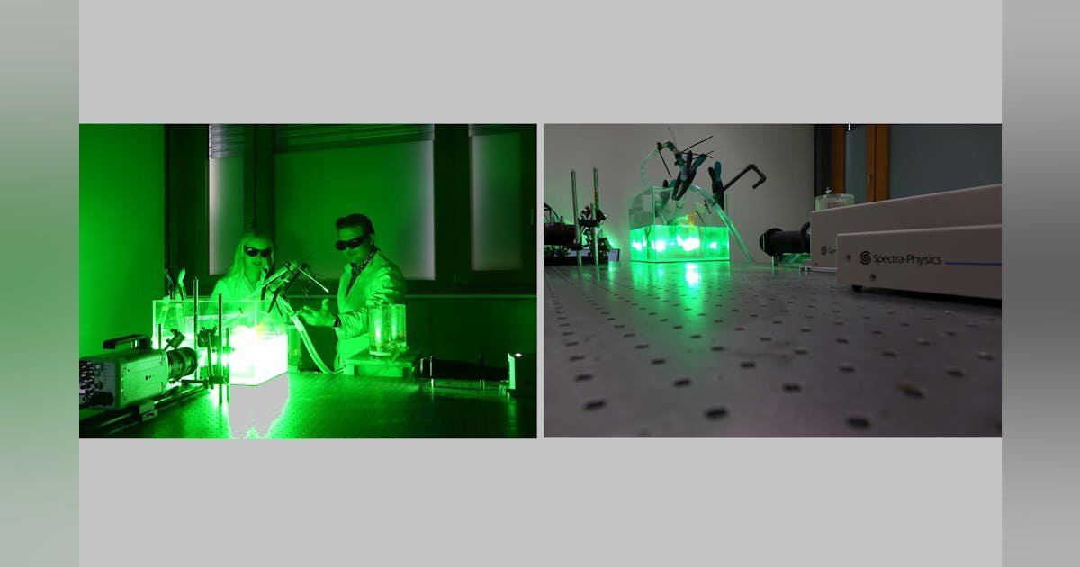
The sophisticated digital twin technology uses “physical” optical measurements as an input. The first step is to make anatomically accurate silicone models based on computer tomography scans of the relevant arteries. To perform realistic in vitro measurements and create a digital twin, the team replicates the relevant in vivo conditions using pulse duplicators.
The experimental optical setup comprises the silicone model, a pulse duplicator to mimic the function of the heart, and the laser system. For the in vitro measurements, the researchers use a fluid mixture composed of glycerin, water, and sodium chloride (NaCl) that mimics the physical properties of blood. Fluorescent rhodamine particles are added to this fluid mixture. And it’s where the laser comes into play: Laser light causes the particles to fluoresce, and a high-resolution camera captures the particle motion. “Illuminating the particles is key to this technology. The pictures not only look artistic but also show the flow within different diseased models and allow us to build and analyze the digital twin,” Guelan explains.