
More ‘pros,’ fewer ‘cons’
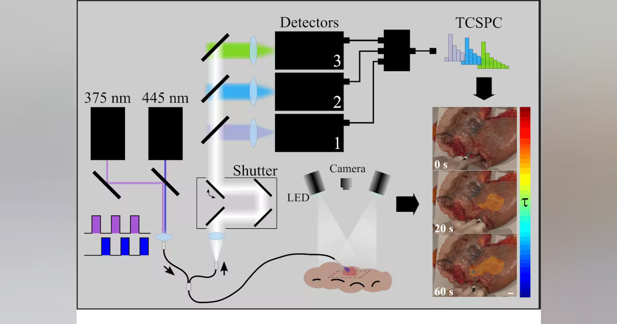
The team’s integrated imaging and analytical approach is label-free, meaning no chemical dyes or contrast agents are needed, which in turn reduces potential patient risks. It also offers real-time analysis, expediting assessment during procedures and aiding precise lesion delineation.
Enhanced diagnostic accuracy is another advantage of the new system, as is the ability to comprehensively characterize tissue by capturing biochemical and structural features that can be further used for evaluating molecular characteristics of the tumor and for outcome prediction.
Lagarto explains there are also several key challenges that exist with current colorectal cancer detection and monitoring methods, which his team’s approach overcomes.
Traditional autofluorescence intensity imaging often suffers from low contrast and is affected by tissue scattering, making it difficult to reliably distinguish between benign and malignant tissues. “By using fluorescence lifetime imaging and phasor-based analysis, our method provides robust, intensity-independent differentiation of tissue types,” Lagarto says.
Another challenge: Standard methods often provide limited functional or molecular information, focusing primarily on morphology. In contrast, the team’s approach delivers comprehensive tissue characterization and captures biochemical and structural features that can inform molecular profiling and outcome prediction, which supports personalized monitoring and treatment planning.
“Beyond surgical oncology, this approach is intended for use during index colonoscopy for early detection,” Lagarto says. “It’s designed for treatment follow-up, as well, which provides a noninvasive tool to monitor disease progression, assess therapy response, and support personalized management strategies.”