
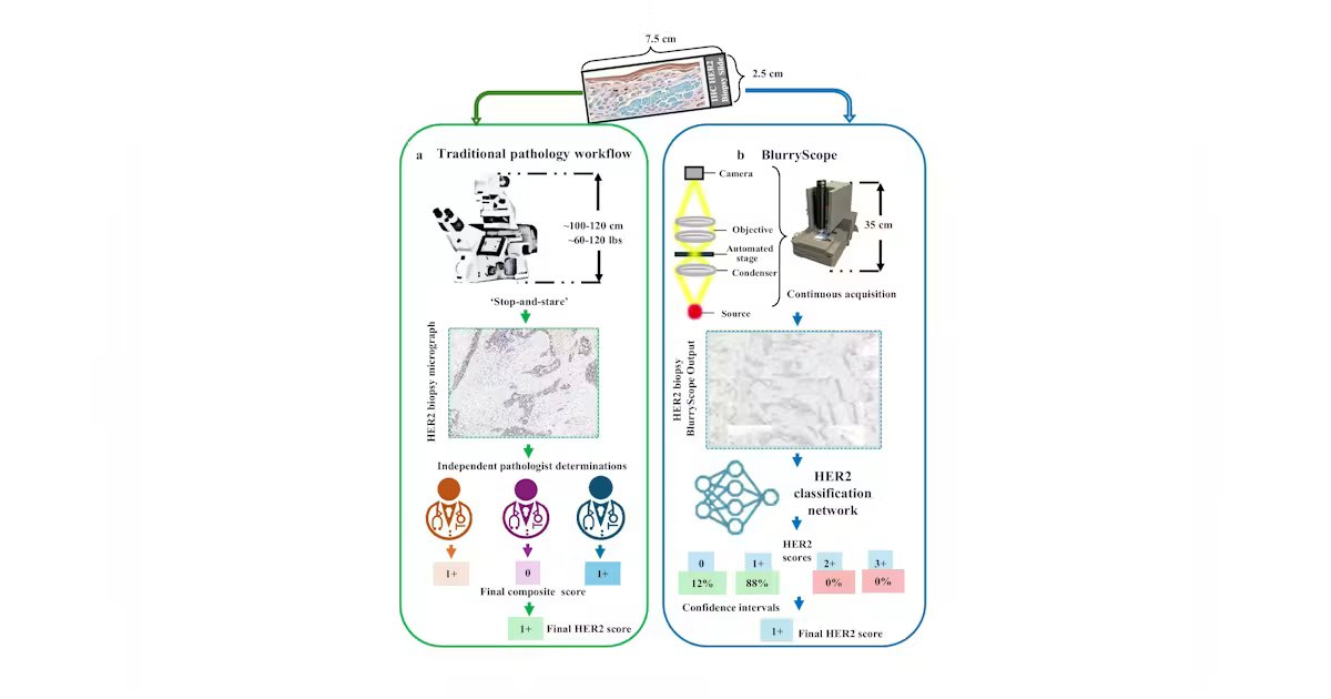
For decades, scanning microscopy has treated motion blur as a defect to be prevented. The familiar stop-and-stare workflow reflects this assumption: Move the specimen, halt the stage, wait for it to settle, expose the camera, and repeat. While it’s a reliable way to protect image quality, the repeated acceleration, settling, and exposure steps also make the mechanics carry much of the system burden.
Computational imaging invites a more adventurous question: If the blur produced by continuous motion is structured and reproducible, can an algorithm recover the information needed for a defined task? It doesn’t make blur desirable in itself, but rather treats it as a precise engineering variable that can be considered alongside stage speed, exposure time, optical resolution, signal-to-noise ratio, training data, and the cost and complexity of the hardware.
Motion blur as a systems-engineering design choice
In earlier work with the late Professor Gabriel Popescu, we tested this idea in GANscan, a continuous-scanning method.1 Rather than stopping the stage for each field of view, the microscope recorded video while the specimen moved. A conditional generative adversarial network (GAN) was trained on registered pairs of motion-blurred and sharp images. Our published experiments used a conventional microscope without specialized scanning hardware and demonstrated restoration at stage speeds up to 5,000 µm per second. In this experimental setting, acquisition throughput was reported at up to 30x the stop-and-stare comparison, while inference on 256-by-256-pixel frames took less than 20 ms on a consumer graphics processing unit (GPU).
Our results established a proof of principle, not a universal permission slip for neural restoration. A network can only be judged against the data, specimen types, optics, motion, and failure modes represented in its validation. The GANscan study compared restored images with slowly scanned and fully stopped controls, evaluated spatial-frequency recovery, and tested tissue from patients outside the training set. It also examined defocus and showed that reconstruction quality declined as the focal offset increased. The limitation is part of the design result: Computation can relax some constraints, but it does not erase physics.
The physics makes the tradeoff concrete. During an exposure, a translating specimen is integrated across the distance traveled by the stage, so the smear grows with velocity and exposure time. At sufficiently high motion, spatial frequencies disappear rather than merely become less contrasty. Classical deconvolution can narrow broadened features, but it can’t recreate information not recorded. A learned model can predict plausible high-frequency structure from examples, which is useful only when that prediction is tested against appropriate controls. For system designers, this means the blur distribution used in training must follow from the actual stage motion, optics, camera integration time, and specimen preparation—not from a generic image-processing recipe.